Ultrasound Imaging
Sonography, or ultrasound as it is commonly known, is a procedure that uses high-frequency sound waves to create an image of the internal structures of your body. Ultrasound is a safe, non-invasive and painless procedure.
What is an ultrasound?
Ultrasound is a safe and painless non-invasive procedure used to image numerous areas in the body to aid in the diagnostic process.
Did you know? Sonography, or ultrasound, can provide a clear picture of soft tissues that don’t show up well on x-ray images.
Ultrasound exams performed at our clinics:
Ultrasounds—being a safe, non-invasive and painless procedure—can be used to image just about any part of the body.
Abdominal
Commonly used to diagnose pain or distention in the upper abdomen, as well as evaluate kidneys, liver, gallbladder, bile ducts, pancreas, and spleen.
Breast
Used to help diagnose breast lumps, or other abnormalities found during a physical exam or screening mammogram.
Cranial/Head
Used to evaluate blood flow to the brain’s major arteries and is often performed on infants whose skulls are not yet completely formed.
Baby Hips
These images help diagnose abnormalities in bone development of the hip.
Intravascular
Helps to identify plaque buildup in the arterial wall—determining your risk for heart attack.
Musculoskeletal (MSK)
Images muscles, tendons, ligaments, nerves and joints throughout your body. Helps in diagnosing sprains, tears, arthritis and other musculoskeletal conditions.
Obstetrical
One of the most common ultrasound procedures that are used to produce a picture of an embryo or fetus within pregnant women, as well as capture an image of the mother’s uterus and ovaries.
Pelvis
Used to image structures and organs in the lower abdomen and pelvis. There are three types of pelvic ultrasound that are used to evaluate reproductive and urinary systems.
Scrotum
Generates an image of a male’s testicles and the surrounding tissue. This procedure helps to evaluate disorders in the testicles, epididymis and scrotum.
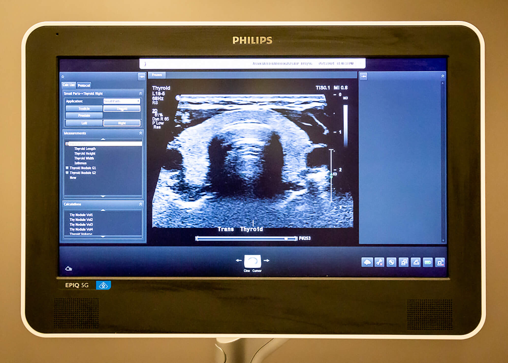
Thyroid
Used to image the thyroid gland in the neck, which helps to regulate a variety of body functions.
Vascular
This procedure helps to identify blockages in the arteries and veins.
How should I prepare for my ultrasound imaging appointment?
Please refer to the ultrasound requisition form provided by your doctor, or the instructions below:
Depending on the type of exam, you may be asked to change into a medical gown.
Abdominal - liver, gallbladder, pancreas, kidneys, and aorta. Do not eat or drink, smoke, chew for 6 hrs before your exam.
Renal and urinary bladder. Drink 1 litre of water 1 hour before your exam and do not empty your bladder prior to your exam.
Abdominal and pelvic. Do not eat for 6 hours before your exam, and drink 1.5 litres of water 1 hour before your exam and do not empty your bladder prior to your exam.
Pelvic - female. Drink 1.5 litres of water 1 hour before your exam and do not empty your bladder prior to your exam.
Pelvic - male. Drink 1 litre of water 1 hour before your exam and do not empty your bladder prior to your exam.
Obstetrical. First 3 months - follow pelvic instructions, above. After 3 months - drink 2 glasses of water 30 minutes before your exam.
What will happen during the sonography procedure?
Most ultrasound exams will have you lying on the exam table, depending on which would result in the highest quality image. The technologist will apply a water-based gel to the area of your body being imaged. The transducer is then moved back and forth over the area until the desired images are captured.


